The isolation of viral DNA – especially from Hepatitis B (HBV) – from serum and plasma has become more important with the growing demands of clinical diagnostics. The extraction of HBV DNA is demanding due to the special genome organization of the Hepatitis B virus. A terminal protein, covalently bound to one of the HBV DNA strands, has to be removed prior to binding of the nucleic acid to the silica membrane. The NucleoSpin® Blood method includes a Proteinase K step for digestion of proteins. Subsequently, the viral DNA is bound to the silica membrane and washed twice with two different buffers ensuring removal of contaminations. Elution can be performed with water or low-salt buffer. The following figures show the outstanding sensitivity of the HBV DNA purification with NucleoSpin® Blood.
Application data
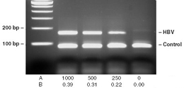
HBV DNA samples purified with NucleoSpin® Blood* were examined using HepScreen B/T amplification assay with integrated fluorescence probe analysis (GENLAB Diagnostics GmbH, Linz/Rh., Germany) Aliquots of each PCR product were characterized by electrophoresis (A), and by endpoint analysis of HBV-specific fluorescence using a microplate fluorometer (B) HBV copies/mL serum (EUROHEP Standard 2) HBV-specific fluorescence (DRHBV)
Purification of HBV DNA with NucleoSpin® Blood* A HBV positive clinical specimen was quantitatively determined by Roche HBV assay. According to the results several dilutions were produced with negative normal serum. In detail, starting with about 500 000 copies HBV/mL, serum samples were diluted down to theoretical about 100 copies/mL. Afterwards, diluted samples were purified with the NucleoSpin® Blood procedure including proteinase K digestion. From 50 μL eluate 5 μL were used for nested PCR amplification procedure. PCR products were analyzed by agarose gel electrophoresis. After nested PCR even highly diluted samples containing only about 100 copies/mL gave positive signals. Positive and negative controls were analyzed in parallel for verification of the test system. Results indicate that NucleoSpin® Virus / Blood procedure shows high recovery rates even for low-titer samples and the purified viral DNA is suitable for appropriate investigations.
Data kindly provided by Dr. C. Tiemann, Laboratory Prof. Hagedorn, Herford, Germany
Quantitative analysis of CMV and EBV from plasma samples Dilutions of the CMV strain AD169 or the EBV Type 1 Strain B95-8 were used as sample. The sample was parallelly diluted into CMV or EBV negative plasma. CMV (red) and EBV (blue) DNA was extracted with NucleoSpin® Blood. The quantitative range using the affigene® CMV trender or affigene® EBV trender (Sangtec Molecular Diagnostics AB, Bromma, Sweden) was analyzed on Mx3000P® (Stratagene, La Jolla, USA). The quantitative range for CMV in plasma was 500–107 c/mL, the range for EBV in plasma was 1000–2 x 10⁷ c/mL.
Data kindly provided by Sangtec Molecular Diagnostics AB, Bromma, Sweden.
Amplification of CMV DNA purified with NucleoSpin® Blood The CMV strain AD169 was serially diluted 1:100 in CMV negative plasma. The CMV DNA was extracted using NucleoSpin® Blood kit, amplified using affigene® CMV trender (Sangtec Molecular Diagnostics AB, Bromma, Sweden), and analyzed on Mx3000P® (Stratagene, La Jolla, USA).
Data kindly provided by Sangtec Molecular Diagnostics AB, Bromma, Sweden.
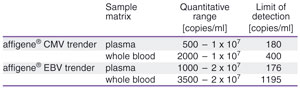
Performance data for CMV and EBV detection using NucleoSpin® Blood for viral DNA extraction Plasma and whole blood were used as sample matrices. CMV and EBV DNA were extracted with NucleoSpin® Blood, and analyzed on Mx3000P® (Stratagene, La Jolla, USA). The table displays the quantitative range and limit of detection (LOD) for the affigene® CMV trender and affigene® EBV trender (Sangtec Molecular Diagnostics AB, Bromma, Sweden).
Data kindly provided by Sangtec Molecular Diagnostics AB, Bromma, Sweden.
The isolation of viral DNA – especially from Hepatitis B (HBV) – from serum and plasma has become more important with the growing demands of clinical diagnostics. The extraction of HBV DNA is demanding due to the special genome organization of the Hepatitis B virus. A terminal protein, covalently bound to one of the HBV DNA strands, has to be removed prior to binding of the nucleic acid to the silica membrane. The NucleoSpin® Blood method includes a Proteinase K step for digestion of proteins. Subsequently, the viral DNA is bound to the silica membrane and washed twice with two different buffers ensuring removal of contaminations. Elution can be performed with water or low-salt buffer. The following figures show the outstanding sensitivity of the HBV DNA purification with NucleoSpin® Blood.
Application data
HBV DNA samples purified with NucleoSpin® Blood* were examined using HepScreen B/T amplification assay with integrated fluorescence probe analysis (GENLAB Diagnostics GmbH, Linz/Rh., Germany) Aliquots of each PCR product were characterized by electrophoresis (A), and by endpoint analysis of HBV-specific fluorescence using a microplate fluorometer (B) HBV copies/mL serum (EUROHEP Standard 2) HBV-specific fluorescence (DRHBV)
Purification of HBV DNA with NucleoSpin® Blood* A HBV positive clinical specimen was quantitatively determined by Roche HBV assay. According to the results several dilutions were produced with negative normal serum. In detail, starting with about 500 000 copies HBV/mL, serum samples were diluted down to theoretical about 100 copies/mL. Afterwards, diluted samples were purified with the NucleoSpin® Blood procedure including proteinase K digestion. From 50 μL eluate 5 μL were used for nested PCR amplification procedure. PCR products were analyzed by agarose gel electrophoresis. After nested PCR even highly diluted samples containing only about 100 copies/mL gave positive signals. Positive and negative controls were analyzed in parallel for verification of the test system. Results indicate that NucleoSpin® Virus / Blood procedure shows high recovery rates even for low-titer samples and the purified viral DNA is suitable for appropriate investigations.
Data kindly provided by Dr. C. Tiemann, Laboratory Prof. Hagedorn, Herford, Germany
Quantitative analysis of CMV and EBV from plasma samples Dilutions of the CMV strain AD169 or the EBV Type 1 Strain B95-8 were used as sample. The sample was parallelly diluted into CMV or EBV negative plasma. CMV (red) and EBV (blue) DNA was extracted with NucleoSpin® Blood. The quantitative range using the affigene® CMV trender or affigene® EBV trender (Sangtec Molecular Diagnostics AB, Bromma, Sweden) was analyzed on Mx3000P® (Stratagene, La Jolla, USA). The quantitative range for CMV in plasma was 500–107 c/mL, the range for EBV in plasma was 1000–2 x 10⁷ c/mL.
Data kindly provided by Sangtec Molecular Diagnostics AB, Bromma, Sweden.
Amplification of CMV DNA purified with NucleoSpin® Blood The CMV strain AD169 was serially diluted 1:100 in CMV negative plasma. The CMV DNA was extracted using NucleoSpin® Blood kit, amplified using affigene® CMV trender (Sangtec Molecular Diagnostics AB, Bromma, Sweden), and analyzed on Mx3000P® (Stratagene, La Jolla, USA).
Data kindly provided by Sangtec Molecular Diagnostics AB, Bromma, Sweden.
Performance data for CMV and EBV detection using NucleoSpin® Blood for viral DNA extraction Plasma and whole blood were used as sample matrices. CMV and EBV DNA were extracted with NucleoSpin® Blood, and analyzed on Mx3000P® (Stratagene, La Jolla, USA). The table displays the quantitative range and limit of detection (LOD) for the affigene® CMV trender and affigene® EBV trender (Sangtec Molecular Diagnostics AB, Bromma, Sweden).
Data kindly provided by Sangtec Molecular Diagnostics AB, Bromma, Sweden.