
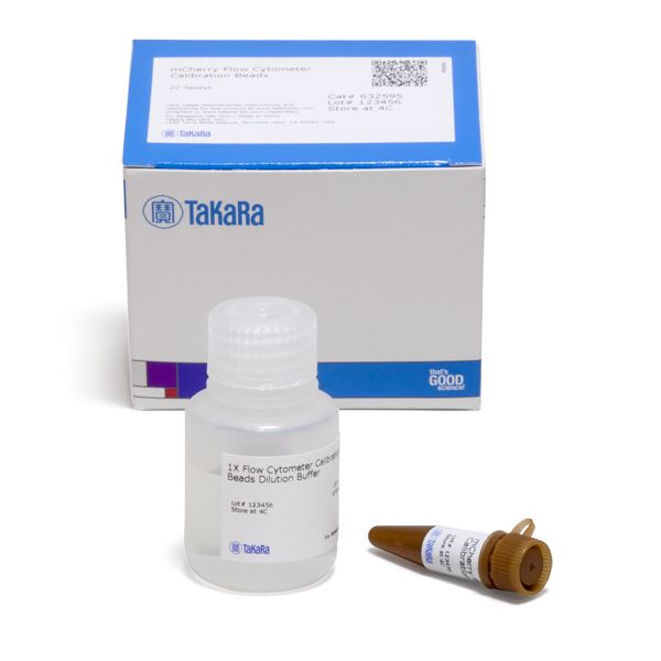
mCherry Flow Cytometer Calibration Beads
mCherry Flow Cytometer Calibration Beads
Brand: Takara Bio.
In stock
SKU
mCherry Flow Cytometer Calibration Beads
The mCherry Flow Cytometer Calibration Beads allow for easy calibration of any flow cytometer with a 561 nm laser line that excites the red fluorescent protein, mCherry. The beads consist of a mixture of six distinct populations that vary in the number of attached mCherry molecules, giving each population a distinct fluorescent signature. The value for the corresponding Molecular Equivalent of Soluble Fluorophore (MESF) per peak was determined by correlating the fluorescence intensity of each respective bead population with the amount of soluble mCherry yielding the same fluorescence intensity. The lowest intensity represents the autofluorescence signal of cells not expressing red fluorescent protein, while the five remaining peaks are evenly distributed over the remaining scale of the red fluorescence detection channel.
Overview
Mixture of six discrete bead populations having distinct fluorescent intensities:
- The lowest intensity represents the autofluorescence signal of cells not expressing the fluorescent protein (AcGFP1 or mCherry)
- The remaining five peaks are evenly distributed over the remaining scale of the green or red fluorescence detection channel
- Works with any flow cytometer with a 488-nm laser line (AcGFP1) or a 561-nm laser line (mCherry)
Applications
- Calibrate your flow cytometer prior to analyzing cells expressing AcGFP1 or mCherry fluorescent protein
- Since the spectral properties and brightness of AcGFP1 and EGFP are very similar, these beads may be used to calibrate flow cytometers prior to using EGFP-expressing cells
Flow cytometer analysis of mCherry Flow Cytometer Calibration Beads

Flow cytometer analysis of mCherry Flow Cytometer Calibration Beads. 20 µl of the mCherry Flow Cytometer Calibration Bead Suspension was thoroughly resuspended in 1ml of 1X Flow Cytometer Calibration Beads Dilution Buffer. The bead suspension was then analyzed on a FACS Diva (BD) flow cytometer using the 561 nm laser excitation line and detecting in the red channel. 10,000 events were analyzed. This graph shows that the bead suspension contains sid well-distinguished populations with different fluorescent intensities.
Write Your Own Review
Flow cytometer analysis of mCherry Flow Cytometer Calibration Beads
Flow cytometer analysis of mCherry Flow Cytometer Calibration Beads. 20 µl of the mCherry Flow Cytometer Calibration Bead Suspension was thoroughly resuspended in 1ml of 1X Flow Cytometer Calibration Beads Dilution Buffer. The bead suspension was then analyzed on a FACS Diva (BD) flow cytometer using the 561 nm laser excitation line and detecting in the red channel. 10,000 events were analyzed. This graph shows that the bead suspension contains sid well-distinguished populations with different fluorescent intensities.