Long ssDNA for Knockins

Long ssDNA for Knockins

Brand: Takara Bio.
In stock
SKU
Long ssDNA for Knockins
Grouped product items
Product Name Size
Guide-it™ Long ssDNA Production System
SKU: 632644
25 Rxns
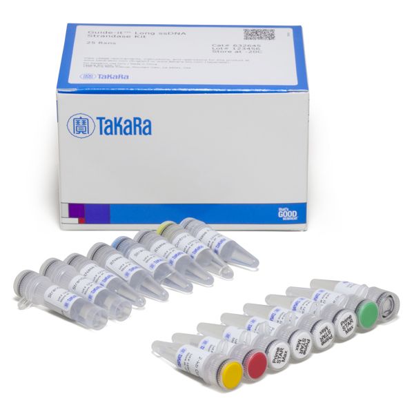
Guide-it™ Long ssDNA Strandase Kit
SKU: 632645
25 Rxns
Make An Enquiry
× Product Enquiry
Long ssDNA for Knockins

Fill in the details below:

The Guide-it Long ssDNA Production System enables production of single-stranded DNA (ssDNA) oligos up to 5 kb in length for use as repair templates in knockin experiments involving CRISPR/Cas9 or other genome editing tools. Included with the system are sufficient reagents for production of 50 different ssDNA oligos (25 pairs of sense and antisense ssDNAs), and the NucleoSpin Gel and PCR Clean-up kit for purification of ssDNA prior to target-cell delivery.

The Guide-it Long ssDNA Strandase Kit enables production of single-stranded DNA (ssDNA) oligos up to 5 kb in length for use as repair templates in knockin experiments involving CRISPR/Cas9 or other genome editing tools. The kit includes sufficient reagents for production of 50 different ssDNA oligos (25 pairs of sense and antisense ssDNAs). In contrast with the Guide-it Long ssDNA Production System (Cat. # 632644), this kit does not include a clean-up kit for purifying ssDNA prior to delivery to target cells.

Overview

  • Create long ssDNA donor templates up to 5 kb in length
  • Fast, easy-to-use, three-step protocol
  • No random integration or expression, leading to improved editing efficiency
  • ssDNA donor templates are far less toxic to cells than dsDNA templates

Applications

  • Donor templates for knockin experiments requiring DNA fragments >500 bp

FACS analysis of AcGFP1 knockin efficiencies at the SEC61B and TUBA1A loci

FACS analysis of AcGFP1 knockin efficiencies at the SEC61B and TUBA1A loci

FACS analysis of AcGFP1 knockin efficiencies at the SEC61B and TUBA1A loci. Cas9-sgRNA RNPs targeting SEC61B and TUBA1A and corresponding AcGFP1 ssDNA homology-directed repair (HDR) templates generated using the Guide-it Long ssDNA Production System were electroporated into ChiPSC18 cells. Following culturing, electroporated target cells and non-electroporated negative control cells were analyzed for GFP expression by FACS. Using this approach, knockin efficiencies of 12.55% and 2.75% were observed for insertion of AcGFP1 at SEC61B and TUBA1A, respectively.

PCR analysis of AcGFP1 knockin at the SEC61B locus in clonal populations isolated by FACS

PCR analysis of AcGFP1 knockin at the SEC61B locus in clonal populations isolated by FACS

PCR analysis of AcGFP1 knockin at the SEC61B locus in clonal populations isolated by FACS. ChiPSC18 cell populations were electroporated with Cas9-sgRNA RNPs and sense or antisense ssDNA HDR templates (labeled ssDNA-1 or ssDNA-2, respectively) that were designed for knockin of AcGFP1 at the SEC61B locus. The cells were sorted on the basis of GFP expression via FACS. Individual clones from the GFP-positive populations were isolated by limiting dilution and cultured to yield clonal cell populations. Genomic DNA extracted from the clonal populations was analyzed by PCR using primers (L-F and R-R, respectively) designed to anneal on either side of the GFP insertion site as shown in the schematic. The GFP and homology arm sequences included in the ssDNA templates are shown in green and light blue, respectively, while the genomic sequences outside the region targeted by the ssDNA homology arms are shown in dark blue. The PCR primers were designed so that the presence of the inserted AcGFP1 sequence yielded a 1.885-kb PCR product, while absence of the AcGFP1 insert yielded a 1.159-kb PCR product. Gel electrophoresis of PCR products confirmed the occurrence of monoallelic knockins of AcGFP1 at SEC61B, represented by double bands (ssDNA-1 Lane C5, and ssDNA-2 Lane C15), and biallelic knockins of AcGFP1 at SEC61B, represented by single bands (ssDNA-1 Lanes C1, C7, C8, C11, and ssDNA-2 Lanes C2, C6, C8, and C10). Two clones lacking any copies of the AcGFP1 insert were also identified (ssDNA-1 Lane C10, and ssDNA-2 Lane C11).

Schematic of Cas9-sgRNA cleavage site and ssDNA construct designed for knockin of AcGFP1 at the TUBA1A locus

Schematic of Cas9-sgRNA cleavage site and ssDNA construct designed for knockin of AcGFP1 at the TUBA1A locus

Schematic of Cas9-sgRNA cleavage site and ssDNA construct designed for knockin of AcGFP1 at the TUBA1A locus. The ssDNA construct generated using the Guide-it Long ssDNA Production System included the AcGFP1 coding sequence (~700 nt in length) flanked on either side by homology arms ~350 nt in length. The homology arms were designed to insert the AcGFP1 sequence at the 5' end of Exon 2 of TUBA1A. As indicated, the ssDNA construct did not include a promoter sequence, so that expression of the resulting AcGFP1-TUBA1A fusion protein would be driven by the endogenous TUBA1A promoter.

Toxicity comparison for electroporation of dsDNA or ssDNA

Toxicity comparison for electroporation of dsDNA or ssDNA

Toxicity comparison for electroporation of dsDNA or ssDNA. ChiPSC18 cells were electroporated with Cas9-sgRNA RNPs targeting the TUBA1A locus, in the absence or presence of homology-directed repair (HDR) templates consisting of dsDNA or ssDNA. After 48 hours of culturing, imaging was performed to compare the toxicity associated with each HDR template. While electroporation of either DNA template resulted in increased toxicity compared to electroporation of Cas9-sgRNA RNPs alone (as evidenced by lower cell counts following culturing), application of the ssDNA template resulted in greatly reduced toxicity compared to the dsDNA template.

In vivo expression of AcGFP1 fusion proteins in cells edited using Cas9-sgRNA RNPs and ssDNA HDR templates

In vivo expression of AcGFP1 fusion proteins in cells edited using Cas9-sgRNA RNPs and ssDNA HDR templates

In vivo expression of AcGFP1 fusion proteins in cells edited using Cas9-sgRNA RNPs and ssDNA HDR templates. ChiPSC18 cells were edited using Cas9-sgRNAs and ssDNA HDR templates designed for knockin of AcGFP1 at the SEC61B and TUBA1A loci, respectively. GFP-positive clonal cell lines obtained from each edited population were then stained with DAPI and analyzed by fluorescence microscopy. Expression of GFP-SEC61B was observed in endosomal compartments (left), while GFP-TUBA1A localized to microtubules (right).

Write Your Own Review
You're reviewing:Long ssDNA for Knockins
Your Rating

The Guide-it Long ssDNA Production System enables production of single-stranded DNA (ssDNA) oligos up to 5 kb in length for use as repair templates in knockin experiments involving CRISPR/Cas9 or other genome editing tools. Included with the system are sufficient reagents for production of 50 different ssDNA oligos (25 pairs of sense and antisense ssDNAs), and the NucleoSpin Gel and PCR Clean-up kit for purification of ssDNA prior to target-cell delivery.

The Guide-it Long ssDNA Strandase Kit enables production of single-stranded DNA (ssDNA) oligos up to 5 kb in length for use as repair templates in knockin experiments involving CRISPR/Cas9 or other genome editing tools. The kit includes sufficient reagents for production of 50 different ssDNA oligos (25 pairs of sense and antisense ssDNAs). In contrast with the Guide-it Long ssDNA Production System (Cat. # 632644), this kit does not include a clean-up kit for purifying ssDNA prior to delivery to target cells.

Overview

  • Create long ssDNA donor templates up to 5 kb in length
  • Fast, easy-to-use, three-step protocol
  • No random integration or expression, leading to improved editing efficiency
  • ssDNA donor templates are far less toxic to cells than dsDNA templates

Applications

  • Donor templates for knockin experiments requiring DNA fragments >500 bp

FACS analysis of AcGFP1 knockin efficiencies at the SEC61B and TUBA1A loci

FACS analysis of AcGFP1 knockin efficiencies at the SEC61B and TUBA1A loci

FACS analysis of AcGFP1 knockin efficiencies at the SEC61B and TUBA1A loci. Cas9-sgRNA RNPs targeting SEC61B and TUBA1A and corresponding AcGFP1 ssDNA homology-directed repair (HDR) templates generated using the Guide-it Long ssDNA Production System were electroporated into ChiPSC18 cells. Following culturing, electroporated target cells and non-electroporated negative control cells were analyzed for GFP expression by FACS. Using this approach, knockin efficiencies of 12.55% and 2.75% were observed for insertion of AcGFP1 at SEC61B and TUBA1A, respectively.

PCR analysis of AcGFP1 knockin at the SEC61B locus in clonal populations isolated by FACS

PCR analysis of AcGFP1 knockin at the SEC61B locus in clonal populations isolated by FACS

PCR analysis of AcGFP1 knockin at the SEC61B locus in clonal populations isolated by FACS. ChiPSC18 cell populations were electroporated with Cas9-sgRNA RNPs and sense or antisense ssDNA HDR templates (labeled ssDNA-1 or ssDNA-2, respectively) that were designed for knockin of AcGFP1 at the SEC61B locus. The cells were sorted on the basis of GFP expression via FACS. Individual clones from the GFP-positive populations were isolated by limiting dilution and cultured to yield clonal cell populations. Genomic DNA extracted from the clonal populations was analyzed by PCR using primers (L-F and R-R, respectively) designed to anneal on either side of the GFP insertion site as shown in the schematic. The GFP and homology arm sequences included in the ssDNA templates are shown in green and light blue, respectively, while the genomic sequences outside the region targeted by the ssDNA homology arms are shown in dark blue. The PCR primers were designed so that the presence of the inserted AcGFP1 sequence yielded a 1.885-kb PCR product, while absence of the AcGFP1 insert yielded a 1.159-kb PCR product. Gel electrophoresis of PCR products confirmed the occurrence of monoallelic knockins of AcGFP1 at SEC61B, represented by double bands (ssDNA-1 Lane C5, and ssDNA-2 Lane C15), and biallelic knockins of AcGFP1 at SEC61B, represented by single bands (ssDNA-1 Lanes C1, C7, C8, C11, and ssDNA-2 Lanes C2, C6, C8, and C10). Two clones lacking any copies of the AcGFP1 insert were also identified (ssDNA-1 Lane C10, and ssDNA-2 Lane C11).

Schematic of Cas9-sgRNA cleavage site and ssDNA construct designed for knockin of AcGFP1 at the TUBA1A locus

Schematic of Cas9-sgRNA cleavage site and ssDNA construct designed for knockin of AcGFP1 at the TUBA1A locus

Schematic of Cas9-sgRNA cleavage site and ssDNA construct designed for knockin of AcGFP1 at the TUBA1A locus. The ssDNA construct generated using the Guide-it Long ssDNA Production System included the AcGFP1 coding sequence (~700 nt in length) flanked on either side by homology arms ~350 nt in length. The homology arms were designed to insert the AcGFP1 sequence at the 5' end of Exon 2 of TUBA1A. As indicated, the ssDNA construct did not include a promoter sequence, so that expression of the resulting AcGFP1-TUBA1A fusion protein would be driven by the endogenous TUBA1A promoter.

Toxicity comparison for electroporation of dsDNA or ssDNA

Toxicity comparison for electroporation of dsDNA or ssDNA

Toxicity comparison for electroporation of dsDNA or ssDNA. ChiPSC18 cells were electroporated with Cas9-sgRNA RNPs targeting the TUBA1A locus, in the absence or presence of homology-directed repair (HDR) templates consisting of dsDNA or ssDNA. After 48 hours of culturing, imaging was performed to compare the toxicity associated with each HDR template. While electroporation of either DNA template resulted in increased toxicity compared to electroporation of Cas9-sgRNA RNPs alone (as evidenced by lower cell counts following culturing), application of the ssDNA template resulted in greatly reduced toxicity compared to the dsDNA template.

In vivo expression of AcGFP1 fusion proteins in cells edited using Cas9-sgRNA RNPs and ssDNA HDR templates

In vivo expression of AcGFP1 fusion proteins in cells edited using Cas9-sgRNA RNPs and ssDNA HDR templates

In vivo expression of AcGFP1 fusion proteins in cells edited using Cas9-sgRNA RNPs and ssDNA HDR templates. ChiPSC18 cells were edited using Cas9-sgRNAs and ssDNA HDR templates designed for knockin of AcGFP1 at the SEC61B and TUBA1A loci, respectively. GFP-positive clonal cell lines obtained from each edited population were then stained with DAPI and analyzed by fluorescence microscopy. Expression of GFP-SEC61B was observed in endosomal compartments (left), while GFP-TUBA1A localized to microtubules (right).
Write Your Own Review
You're reviewing:Long ssDNA for Knockins
Your Rating