
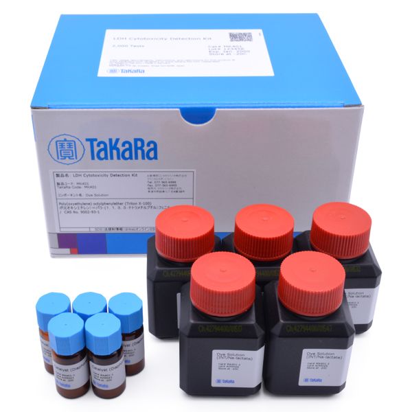
LDH Cytotoxicity Detection Kit
LDH Cytotoxicity Detection Kit
Brand: Takara Bio.
In stock
SKU
LDH Cytotoxicity Detection Kit
The LDH Cytotoxicity Detection Kit uses LDH, a stable cytoplasmic enzyme that is present in large amounts in most cells. LDH is released into the cell culture supernatant during cytoplasmic membrane damage and can be easily measured using standard reagents.
Overview
- Safety: No radioactive isotopes required
- Accuracy: Assay results strongly correlate with the number of damaged cells
- High sensitivity: Low cell numbers (e.g., 0.2–2 x 10² cells/well) are easily detected
- Fast: Only 0.5–1 hour required for measurement
- Simple: Relabeling and washing steps not required. Radiation disposal and safety procedures not required
Applications
- Detection of lactate dehydrogenase (LDH, LD) released from damaged cells
- LDH cytotoxicity detection
LDH assay confirms that cell death in the presence of 1% Triton X-100 is proportional to the number of cells in the well

LDH assay confirms that cell death in the presence of 1% Triton X-100 is proportional to the number of cells in the well. K562 cells were titrated in 96-well plates at the indicated cell concentrations. The reaction mixture was added to confirm that the spontaneous release of LDH is low. In the presence of the reaction mixture plus Triton X-100 (final concentration 1%), LDH release increased proportionally to cell concentration, based on measurements of formazan absorbance at 492 nm.
Using the LDH assay to measure the cytolytic activity of allogen-stimulated, cytotoxic T lymphocytes

Using the LDH assay to measure the cytolytic activity of allogen-stimulated, cytotoxic T lymphocytes. Spleen cells of C57/Bl 6 mice (H-2b) were stimulated in vitro with P815 cells (H-2d). Viable cytotoxic T lymphocytes (CTLs) were purified by ficoll density gradient, washed and titrated in a 96-well plate. 1 × 10⁴ P815 test cells/well were added to the effector CTL cells. The cell mixture was centrifuged and incubated for 4 hr. 100 μl of culture supernatant was collected for the LDH assay. Panel A. Absorbance values for the effector cell control (orange), effector-test cell mix (blue), and effector-test cell mix minus effector cell control (red). Panel B. Percent cell-mediated cytotoxicity.
Principle of measurement of LDH cytotoxicity

Principle of measurement of LDH cytotoxicity.
Baba, H. et al. Induction of gamma interferon and nitric oxide by truncated pneumolysin that lacks pore-forming activity. Infect. Immun. 70, 107–13 (2002).
Ito, Y. et al. Seeligeriolysin O, a cholesterol-dependent cytolysin of Listeria seeligeri, induces gamma interferon from spleen cells of mice. Infect. Immun. 71, 234–41 (2003).
Kikuchi, T. et al. Tumor suppression induced by intratumor administration of adenovirus vector expressing NK4, a 4-kringle antagonist of hepatocyte growth factor, and naive dendritic cells. Blood 100, 3950–9 (2002).
Kohda, C. et al. Dissociated linkage of cytokine-inducing activity and cytotoxicity to different domains of listeriolysin O from Listeria monocytogenes. Infect. Immun. 70, 1334–41 (2002).
Lee, M.-J. et al. Bone morphogenetic protein-7 inhibits constitutive and interleukin-1 beta-induced monocyte chemoattractant protein-1 expression in human mesangial cells: role for JNK/AP-1 pathway. J. Immunol. 170, 2557–63 (2003).
Noda, M. et al. Switch to anaerobic glucose metabolism with NADH accumulation in the beta-cell model of mitochondrial diabetes. Characteristics of betaHC9 cells deficient in mitochondrial DNA transcription. J. Biol. Chem. 277, 41817–26 (2002).
Tabrizi, S. J. et al. Expression of mutant alpha-synuclein causes increased susceptibility to dopamine toxicity. Hum. Mol. Genet. 9, 2683–9 (2000).
Tsuchiya, K. et al. Listeriolysin O-induced membrane permeation mediates persistent interleukin-6 production in Caco-2 cells during Listeria monocytogenes infection in vitro. Infect. Immun. 73, 3869–77 (2005).
Tsukahara, T. et al. Identification of human autologous cytotoxic T-lymphocyte-defined osteosarcoma gene that encodes a transcriptional regulator, papillomavirus binding factor. Cancer Res. 64, 5442–8 (2004).
Zhang, H. M., Ou, Z. L. & Yamamoto, T. Anisodamine inhibits shiga toxin type 2-mediated tumor necrosis factor-alpha production in vitro and in vivo. Exp. Biol. Med. (Maywood). 226, 597–604 (2001).
Write Your Own Review
LDH assay confirms that cell death in the presence of 1% Triton X-100 is proportional to the number of cells in the well
LDH assay confirms that cell death in the presence of 1% Triton X-100 is proportional to the number of cells in the well. K562 cells were titrated in 96-well plates at the indicated cell concentrations. The reaction mixture was added to confirm that the spontaneous release of LDH is low. In the presence of the reaction mixture plus Triton X-100 (final concentration 1%), LDH release increased proportionally to cell concentration, based on measurements of formazan absorbance at 492 nm.
Using the LDH assay to measure the cytolytic activity of allogen-stimulated, cytotoxic T lymphocytes
Using the LDH assay to measure the cytolytic activity of allogen-stimulated, cytotoxic T lymphocytes. Spleen cells of C57/Bl 6 mice (H-2b) were stimulated in vitro with P815 cells (H-2d). Viable cytotoxic T lymphocytes (CTLs) were purified by ficoll density gradient, washed and titrated in a 96-well plate. 1 × 10⁴ P815 test cells/well were added to the effector CTL cells. The cell mixture was centrifuged and incubated for 4 hr. 100 μl of culture supernatant was collected for the LDH assay. Panel A. Absorbance values for the effector cell control (orange), effector-test cell mix (blue), and effector-test cell mix minus effector cell control (red). Panel B. Percent cell-mediated cytotoxicity.
Principle of measurement of LDH cytotoxicity
Principle of measurement of LDH cytotoxicity.