
In Situ Apoptosis Detection Kit
In Situ Apoptosis Detection Kit
Brand: Takara Bio.
In stock
SKU
In Situ Apoptosis Detection Kit
This kit is designed to detect fragmented DNA histochemically by terminal labeling. TUNEL method (Terminal deoxynucleotidyl transferase-mediated dUTP nick end labeling) is an effective method for measuring the DNA fragments resulting from the apoptotic activation of intracellular endonucleases. Fluorescein labeled nucleotides are in situ incorporated onto the ends of these DNA fragments, allowing histologic localization and individual cells to be detected.
Overview
- Quick and easy detection of apoptotic cells
- Sensitive detection of single cells in the initial stages of apoptosis
- Specific staining of apoptotic but not necrotic cells
- Flexible enough to be used with tissue section or fixed-cell samples
- Individual components are available separately
- Control slide supplied for accuracy
- Non-radioactive
Applications
- Fragmented-DNA detection by the TUNEL method
- Detection of DNA strand breaks in apoptotic cells via fluorescence or light microscopy
Fragmented DNA detection by the TUNEL method
Type: direct TUNEL labeling assay
Used for: detection of DNA strand breaks in apoptotic cells by flow cytometry or fluorescence microscopy
Used to assay: cells in suspension (permanent cell lines, normal and tumor cells ex vivo), adherent cells, cytospins, cell smears, frozen or paraffin-embedded tissue sections
Technique: end-labeling of DNA with fluorescein-dUTP, followed by direct analysis of fluorescent cells
Time required: 1–2 hr (Not including sample preparation, permeabilization, etc.)
Detection method: fluorescence and light microscopy
Number of Assays per Kit: 20
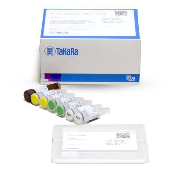
Kit components (Cat. # MK500)
- Labeling Safe Buffer (2 x 500 ml)
- TdT (terminal deoxynucleotidyl transferase) enzyme* (2 x 50 ml)
- Anti-FITC HRP Conjugate (1.5 ml)
- Control Slides (2 slides)
- Permeabilization buffer (2 x 1.0 ml)
*Source: Recombinant E. coli encoding the TdT gene
**Control slides contain paraffin-embedded tissue sections of rat mammary gland. When used as a positive control, deparaffinization is required. Following deparaffinization and treatment with proteinase K, please follow the listed detection method protocol.
Storage
- Shipped at –20°C.
- Components should be stored separately as follows: Labeling Safe Buffer, TdT enzyme, and Permeabilization buffer at –20°C; Anti-FITC HRP Conjugate at 4°C* and Control slides at room temperature.**
*Store at 4°C when thawed.
**Store at room temperature after delivery; kit is shipped at –20°C.
Write Your Own Review