
Fluorescent Transfection Controls
Fluorescent Transfection Controls
Brand: Takara Bio.
In stock
SKU
Fluorescent Transfection Controls
Fluorescent labeling and transfection control
These vectors are ideally suited as controls to measure transfection efficiency. Simply co-transfect a red fluorescent protein vector with the target plasmid, and the resulting cells that are labeled with the fluorescent protein will also likely be transfected with the experimental plasmid. Each vector expresses a fluorescent protein from a constitutive promoter (CMV or EF1-alpha) and, if required, stable transfected cells can be selected for using G418. Transfected cells can also be used in assays that require fluorescent labeling of whole cells.
These vectors do not contain a multiple cloning site; other fluorescent protein vectors are available for cloning applications. Click here for green fluorescent protein options.
Overview
- Bright fluorescent proteins for labeling transfected cells
- Choice of CMV or EF1-alpha promoter
- CMV promoter for the strongest transcriptional activity in the broadest range of cell types
- Human elongation factor-1 alpha (EF1-alpha) promoter that can be used in cells where CMV can be silenced (e.g., embryonic stem cells and some hematopoietic cells)
- Multiple red color options
- tdTomato is the brightest of the red fluorescent proteins and is also extremely suitable for in vivo imaging applications
- E2-Crimson, a far-red fluorescent protein, is bright, very soluble, and matures extremely fast (half time of 26 minutes at 37°C). It exhibits low cytotoxicity and is well-suited for in vivo applications involving sensitive cells such as primary cells and stem cells.
Applications
- Fluorescent reporter for transient and stable transfections
- Whole-cell labeling
Robust expression of DsRed-Express2 in mouse bone marrow hematopoietic stem and progenitor cells
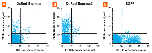
Robust expression of DsRed-Express and DsRed-Express2 in mouse bone marrow hematopoietic stem and progenitor cells. Mononuclear bone marrow cells were transduced with retroviral vectors encoding DsRed-Express (Panel A), DsRed-Express2 (Panel B), or EGFP (Panel C), and fluorescent cells were sorted 87 hr later. Red and green fluorescence signals were detected using the PE and FITC filter sets, respectively. The lines represent gates defined by analyzing untransduced cells.
tdTomato, but not GFP, can be detected in the SCID mouse cadaver phantom model
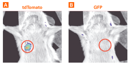
tdTomato, but not GFP, can be detected in the SCID mouse cadaver phantom model. False-color overlay images (regions of interest encircled) indicate that the imaging system could detect tdTomato fluorescence in the cadaver model, but not GFP fluorescence. Panel A. Implanted tube with 100 x 106 MDA-MB-231-tdTomato-expressing cells, imaged with the DsRed filter set. Exposure time: 1 sec. Panel B. Implanted tube with 100 x 106 MDA-MB-231-GFP-expressing cells, imaged with the GFP filter set. Exposure time: 1 sec.
Map schematic of the plasmid choices that are available carrying the EF1-alpha promoter
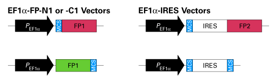
Map schematic of the plasmid choices that are available carrying the EF1-alpha promoter. IRES: internal ribosome entry sequence; FP1: fluorescent protein (AcGFP1, DsRed-Monomer, or mCherry); FP2: fluorescent protein (mCherry or ZsGreen1); MCS: multiple cloning site.
Write Your Own Review
Robust expression of DsRed-Express2 in mouse bone marrow hematopoietic stem and progenitor cells
Robust expression of DsRed-Express and DsRed-Express2 in mouse bone marrow hematopoietic stem and progenitor cells. Mononuclear bone marrow cells were transduced with retroviral vectors encoding DsRed-Express (Panel A), DsRed-Express2 (Panel B), or EGFP (Panel C), and fluorescent cells were sorted 87 hr later. Red and green fluorescence signals were detected using the PE and FITC filter sets, respectively. The lines represent gates defined by analyzing untransduced cells.
tdTomato, but not GFP, can be detected in the SCID mouse cadaver phantom model
tdTomato, but not GFP, can be detected in the SCID mouse cadaver phantom model. False-color overlay images (regions of interest encircled) indicate that the imaging system could detect tdTomato fluorescence in the cadaver model, but not GFP fluorescence. Panel A. Implanted tube with 100 x 106 MDA-MB-231-tdTomato-expressing cells, imaged with the DsRed filter set. Exposure time: 1 sec. Panel B. Implanted tube with 100 x 106 MDA-MB-231-GFP-expressing cells, imaged with the GFP filter set. Exposure time: 1 sec.
Map schematic of the plasmid choices that are available carrying the EF1-alpha promoter
Map schematic of the plasmid choices that are available carrying the EF1-alpha promoter. IRES: internal ribosome entry sequence; FP1: fluorescent protein (AcGFP1, DsRed-Monomer, or mCherry); FP2: fluorescent protein (mCherry or ZsGreen1); MCS: multiple cloning site.