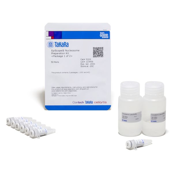
EpiScope Nucleosome Preparation Kit

EpiScope Nucleosome Preparation Kit

Brand: Takara Bio.
In stock
SKU
EpiScope Nucleosome Preparation Kit
Grouped product items
Product Name Size
EpiScope® Nucleosome Preparation Kit
SKU: 5333
50 Rxns
Make An Enquiry
× Product Enquiry
EpiScope Nucleosome Preparation Kit

Fill in the details below:

This kit is designed for the preparation of nucleosomes from cultured mammalian cells. DNA can be extracted from the prepared nucleosome and analyzed using a real-time PCR assay or high-speed sequencing to map nucleosome positions.

Overview

  • Enables detailed analysis of nucleosome positioning
  • Prepared DNA consists primarily of mononucleosomes

Applications

  • Preparation of nucleosomal DNA from 0.5–2 x 10⁶ mammalian cultured cells
  • Nucleosome positioning with regards to DNA methylation analysis

Genomic DNA regions occupied with nucleosomes are quantified or identified with downstream applications, such as qPCR, DNA sequencing, and microarray

Genomic DNA regions occupied with nucleosomes are quantified or identified with downstream applications, such as qPCR, DNA sequencing, and microarray

Genomic DNA regions occupied with nucleosomes are quantified or identified with downstream applications, such as qPCR, DNA sequencing, and microarray.

Protocol overview

Protocol overview

Protocol overview.

Analysis of nucleosomal DNA size distribution

Analysis of nucleosomal DNA size distribution

Analysis of nucleosomal DNA size distribution. Nucleosomal DNA preparation from HeLa cells (1 x 10⁶ cells) was performed with various amounts of micrococcal nuclease (0.02 , 0.05, and 0.2 ul). Nucleosomal DNA was purified with the NucleoSpin Extract II, yielding 50 ul of DNA solution, of which 5 ul was analyzed by agarose gel electrophoresis. For samples treated with lower amounts of micrococcal nuclease (0.02 ul, 0.05 ul), the electrophoretic results showed bands for mononucleosomal, dinucleosomal, and trinucleosomal DNA fragments. For samples treated with 0.2 ul of micrococcal nuclease, mostly mononucleosomes were evident. In addition, the higher the amount of micrococcal nuclease used, the shorter the size of the mononucleosome-derived DNA fragment observed. This result may reflect the trimming activity at both ends of the DNA (Clark et al. 2010).

Clark, D. J. Nucleosome positioning, nucleosome spacing and the nucleosome code. J. Biomol. Struct. Dyn. 27, 781–93 (2010).   

Dutta, A. & Workman, J. L. Nucleosome Positioning: Multiple Mechanisms toward a Unifying Goal. Mol. Cell 48, 1–2 (2012).

Write Your Own Review
You're reviewing:EpiScope Nucleosome Preparation Kit
Your Rating

This kit is designed for the preparation of nucleosomes from cultured mammalian cells. DNA can be extracted from the prepared nucleosome and analyzed using a real-time PCR assay or high-speed sequencing to map nucleosome positions.

Overview

  • Enables detailed analysis of nucleosome positioning
  • Prepared DNA consists primarily of mononucleosomes

Applications

  • Preparation of nucleosomal DNA from 0.5–2 x 10⁶ mammalian cultured cells
  • Nucleosome positioning with regards to DNA methylation analysis

Genomic DNA regions occupied with nucleosomes are quantified or identified with downstream applications, such as qPCR, DNA sequencing, and microarray

Genomic DNA regions occupied with nucleosomes are quantified or identified with downstream applications, such as qPCR, DNA sequencing, and microarray

Genomic DNA regions occupied with nucleosomes are quantified or identified with downstream applications, such as qPCR, DNA sequencing, and microarray.

Protocol overview

Protocol overview

Protocol overview.

Analysis of nucleosomal DNA size distribution

Analysis of nucleosomal DNA size distribution

Analysis of nucleosomal DNA size distribution. Nucleosomal DNA preparation from HeLa cells (1 x 10⁶ cells) was performed with various amounts of micrococcal nuclease (0.02 , 0.05, and 0.2 ul). Nucleosomal DNA was purified with the NucleoSpin Extract II, yielding 50 ul of DNA solution, of which 5 ul was analyzed by agarose gel electrophoresis. For samples treated with lower amounts of micrococcal nuclease (0.02 ul, 0.05 ul), the electrophoretic results showed bands for mononucleosomal, dinucleosomal, and trinucleosomal DNA fragments. For samples treated with 0.2 ul of micrococcal nuclease, mostly mononucleosomes were evident. In addition, the higher the amount of micrococcal nuclease used, the shorter the size of the mononucleosome-derived DNA fragment observed. This result may reflect the trimming activity at both ends of the DNA (Clark et al. 2010).

Clark, D. J. Nucleosome positioning, nucleosome spacing and the nucleosome code. J. Biomol. Struct. Dyn. 27, 781–93 (2010).   

Dutta, A. & Workman, J. L. Nucleosome Positioning: Multiple Mechanisms toward a Unifying Goal. Mol. Cell 48, 1–2 (2012).

Write Your Own Review
You're reviewing:EpiScope Nucleosome Preparation Kit
Your Rating