
CERO 3D Cultivation System
CERO 3D Cultivation System
Brand: OMNI Life Science
In stock
SKU
CERO 3D Cultivation System
Think in new dimensions – CERO is overcoming known limitations of static cultures. Easy, ready-to-use protocols, standardizable workflows and autoadhesion of cells without any need for Carriers or Matrigel are just a few of the advantages. As a result, CERO enables high yield expansion of pluripotent stem cells or long term cultivation of tissue for >20 days (Spheroids >80 or Organoids >180).
Highlights
- Reduces cost, time spend and variations
- Improved viability and maturation
- Significantly reduced apoptosis & necrosis
- Homogenous condition for homogeneous results
- No shear forces and minimized gravity
- Enabling pathogenic infection studies
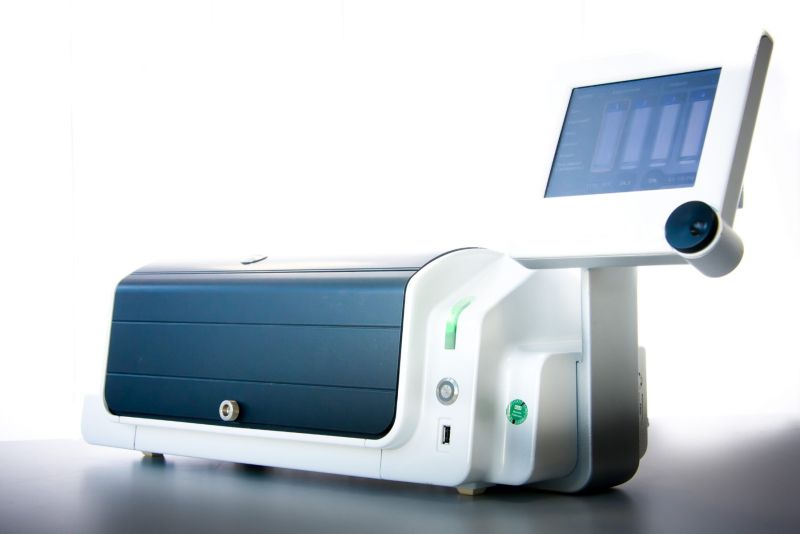
CERO. A Benchtop Incubator & Bioreactor
Convenient, Simple Process
High cell yields, long term culture, cost-effective
Relevant Cells
Proprietary 3D cell culture technology, better diffusion, accelerated maturation
Consistent Results
Scalable to production level, automatable
Outstanding Cell Quality
Fully functional and validated cells
Less Workload
Time-saving, automatic procedures, standardised protocols, walk-away operation
Highest Yield
More mature cells, more relevant cells for drug discovery
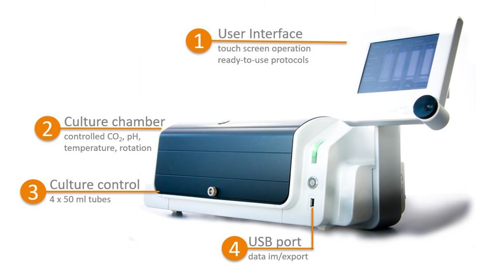
Description of the CERO benchtop bioreactor. CERO is a benchtop incubator and bioreactor hybrid with no impellers for micro-carrier and suspension cell culture. Most of the existing products require large media volumes, complex software or non-intuitive preparation of bioreactor equipment predominantly suited for large scale production. Increasingly, pluripotent stem cells and organoids as standard tools for basic research are implemented for drug development and predictive toxicity screen.
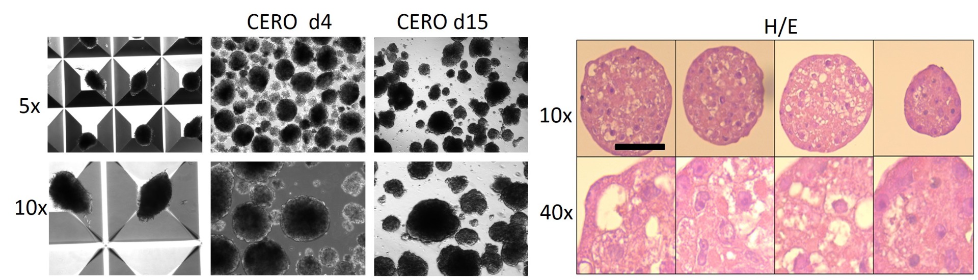
Generation of spheroids derived from HepaRG cells. (left image) Macroscopic analyses of CERO and Aggrewell-induced spheroids. (right image) Histological analyses by H/E of various HepaRG derived sperhoids - used for viral expression analyses. Scale bar: 100mm.
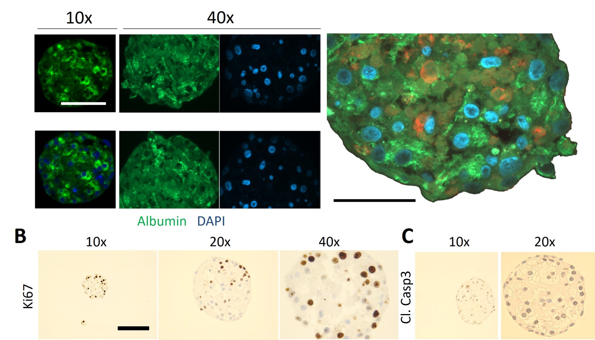
Charatcerization of spheroids derived from HepaRG cells. (left image) (A-C) Microscopic analyses of CERO induced spheroids analysed for albumin and cytokeratin expression, proliferation (Ki67) and cell death (Cl.Casp3). Scale bars: 100mm.
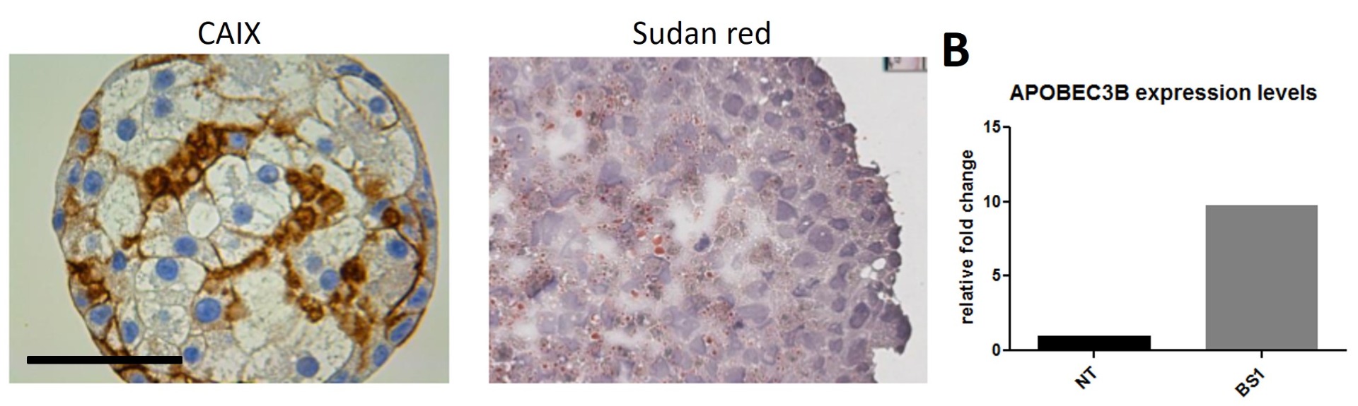
Characterization of spheroids derived from HepaRG cells. (A, left image) Expression of the hypoxia-marker-CAIX in spheroids. (A, right image) Identification of lipids on cryo sections by sudan red. Macroscopic analyses of CERO and Aggrewell induced spheroids. (C) Expression analysis of the cell intrinsic anti-HBV factor APOBEC3B upon stimulation with a LTbR-agonist (BS1) (Lucifora et al., Science 2014). Scale bar: 100mm.
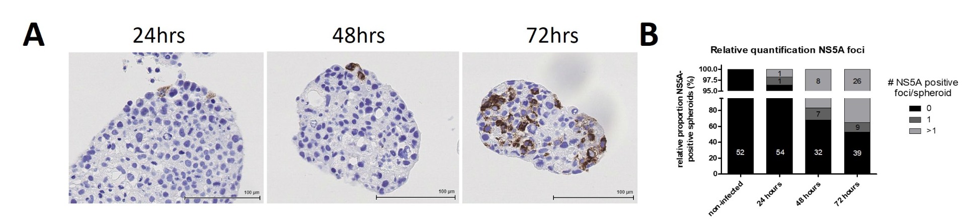
HCV replication in Huh7 derived spheroids over time. (A) Analyses for HCV- replication over time (24-72hrs). (B) Quantification of the overall infection experiment over time. Size of scale bars is indicated.
Write Your Own Review
Description of the CERO benchtop bioreactor. CERO is a benchtop incubator and bioreactor hybrid with no impellers for micro-carrier and suspension cell culture. Most of the existing products require large media volumes, complex software or non-intuitive preparation of bioreactor equipment predominantly suited for large scale production. Increasingly, pluripotent stem cells and organoids as standard tools for basic research are implemented for drug development and predictive toxicity screen.
Generation of spheroids derived from HepaRG cells. (left image) Macroscopic analyses of CERO and Aggrewell-induced spheroids. (right image) Histological analyses by H/E of various HepaRG derived sperhoids - used for viral expression analyses. Scale bar: 100mm.
Charatcerization of spheroids derived from HepaRG cells. (left image) (A-C) Microscopic analyses of CERO induced spheroids analysed for albumin and cytokeratin expression, proliferation (Ki67) and cell death (Cl.Casp3). Scale bars: 100mm.
Characterization of spheroids derived from HepaRG cells. (A, left image) Expression of the hypoxia-marker-CAIX in spheroids. (A, right image) Identification of lipids on cryo sections by sudan red. Macroscopic analyses of CERO and Aggrewell induced spheroids. (C) Expression analysis of the cell intrinsic anti-HBV factor APOBEC3B upon stimulation with a LTbR-agonist (BS1) (Lucifora et al., Science 2014). Scale bar: 100mm.
HCV replication in Huh7 derived spheroids over time. (A) Analyses for HCV- replication over time (24-72hrs). (B) Quantification of the overall infection experiment over time. Size of scale bars is indicated.