
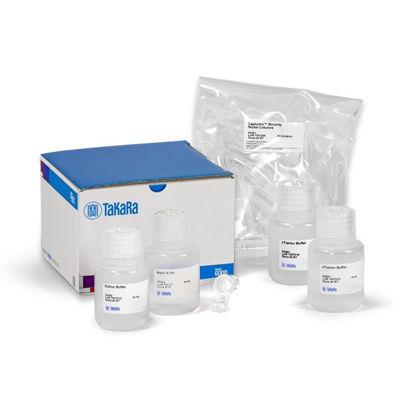
Capturem His-Tagged Purification Miniprep Kit
Capturem His-Tagged Purification Miniprep Kit
Brand: Takara Bio.
In stock
SKU
Capturem His-Tagged Purification Miniprep Kit
Capturem high-capacity membranes
The Capturem His-Tagged Purification Miniprep Kit provides single-use disposable membrane spin columns for simple, rapid purification of his-tagged proteins in up to 800 μl of clarified lysate. The columns are suitable for use under native or denaturing conditions, in the presence of additives such as DTT (up to 10 mM), βME (up to 30 mM), TCEP (up to 5 mM), EDTA (up to 10 mM), or glycerol.
Overview
- No-waiting workflow—5-minute, room-temperature protocol
- High purity and yield—small column bed volumes trap fewer contaminants and allow large-volume washes
- Compatible with wide range of conditions—native and denaturing conditions; with common additives (e.g., EDTA, DTT, BME, glycerol, TCEP, etc.; see compatibility table)
- Lysis buffer compatibility—xTractor Buffer; buffers from other vendors
- Different cell systems—easy purification from mammalian and bacterial cells
Applications
- Biochemical/enzymatic assays
- Pull-down or other protein interaction assays
- Confirmation screening
- Protein/ligand affinity analysis
- Crystallography or other structural analyses
- Antibody production
- Toxicology studies
- Protein transfer to live cells
Protein purification miniprep workflow

Protein purification miniprep workflow. Each mini spin column can be loaded with up to 800 μl of lysate (yielded from 2–5 ml of culture). His-tagged protein is first bound to the membrane, followed by washing with 300 μl wash buffer, and elution with 300 μl elution buffer. Over 90% of the bound protein can be eluted with as little as 100 μl elution buffer. Each step is followed by spinning the tube for 1 min at 11,000 x g. The working bed volume of the membrane is <2 μl. This entire purification is complete in <5 min.
Purification of GFPuv in the presence of a wide range of additives, at varying concentrations

Purification of GFPuv in the presence of a wide range of additives, at varying concentrations. Additives were included in the sample, equilibration, wash, and elution buffers for each experiment. Samples were eluted twice with 300 µl elution buffer each time.
Purification of GFPuv under native and denaturing conditions

Purification of GFPuv under native and denaturing conditions. Spin columns were loaded with 800 µl of cell lysate, and all steps were performed with appropriate buffers for native and denaturing conditions. 8 M urea or 6 M guanidine were included in appropriate samples.
Purification of 6xhis-tagged proteins expressed in mammalian cells

Purification of 6xhis-tagged proteins expressed in mammalian cells. 293T cells were transfected with plasmids encoding 6xhis-tagged MAPK1 or ADRB2. Cells were lysed, and the clarified lysate was loaded onto an equilibrated column. The column was washed with 400 μl wash buffer, followed by elution with 300 μl elution buffer. The various fractions were then resolved on a 4–20% polyacrylamide gel, and blotted onto a nitrocellulose membrane. Panel A, left. Ponceau S staining of the MAPK1 membrane shows all the proteins present in the different fractions. Panel A, right. Immunoblot using an antibody specific to MAPK1 shows a band corresponding to MAPK1. Panel B, left. Ponceau S staining of the ADRB2 membrane shows all the proteins present in the different fractions. Panel B, right. Immunoblot using an antibody specific to ADRB2 shows the enrichment of ADRB2 in the eluate.
Capturem Protein A- or G-functionalized membranes

Antibody purification workflow for Capturem Protein A or G 96-well plates. Each well can be loaded with up to 850 µl of a diluted sample (antibody sample diluted from 1:1 to 1:20 with buffer). Following binding, the membrane is then washed and the bound antibiody eluted with the appropriate buffers. Note: Protocol time varies for each format. Check user manuals for specifics on protocol times.
Capturem spin columns and filtration devices

Available Capturem spin column and filtration device formats. Pictured from left to right: nanoprep, miniprep, 96-well, 24-well, maxiprep, and large-volume Capturem formats. Formats available vary for the different functionalized membranes. Check product pages for list of available formats.
Load volumes and approximate yields for available Capturem formats by chemistry

Load volumes and yields of Capturem purification formats.
Write Your Own Review
Protein purification miniprep workflow
Protein purification miniprep workflow. Each mini spin column can be loaded with up to 800 μl of lysate (yielded from 2–5 ml of culture). His-tagged protein is first bound to the membrane, followed by washing with 300 μl wash buffer, and elution with 300 μl elution buffer. Over 90% of the bound protein can be eluted with as little as 100 μl elution buffer. Each step is followed by spinning the tube for 1 min at 11,000 x g. The working bed volume of the membrane is <2 μl. This entire purification is complete in <5 min.
Purification of GFPuv in the presence of a wide range of additives, at varying concentrations
Purification of GFPuv in the presence of a wide range of additives, at varying concentrations. Additives were included in the sample, equilibration, wash, and elution buffers for each experiment. Samples were eluted twice with 300 µl elution buffer each time.
Purification of GFPuv under native and denaturing conditions
Purification of GFPuv under native and denaturing conditions. Spin columns were loaded with 800 µl of cell lysate, and all steps were performed with appropriate buffers for native and denaturing conditions. 8 M urea or 6 M guanidine were included in appropriate samples.
Purification of 6xhis-tagged proteins expressed in mammalian cells
Purification of 6xhis-tagged proteins expressed in mammalian cells. 293T cells were transfected with plasmids encoding 6xhis-tagged MAPK1 or ADRB2. Cells were lysed, and the clarified lysate was loaded onto an equilibrated column. The column was washed with 400 μl wash buffer, followed by elution with 300 μl elution buffer. The various fractions were then resolved on a 4–20% polyacrylamide gel, and blotted onto a nitrocellulose membrane. Panel A, left. Ponceau S staining of the MAPK1 membrane shows all the proteins present in the different fractions. Panel A, right. Immunoblot using an antibody specific to MAPK1 shows a band corresponding to MAPK1. Panel B, left. Ponceau S staining of the ADRB2 membrane shows all the proteins present in the different fractions. Panel B, right. Immunoblot using an antibody specific to ADRB2 shows the enrichment of ADRB2 in the eluate.
Capturem Protein A- or G-functionalized membranes
Antibody purification workflow for Capturem Protein A or G 96-well plates. Each well can be loaded with up to 850 µl of a diluted sample (antibody sample diluted from 1:1 to 1:20 with buffer). Following binding, the membrane is then washed and the bound antibiody eluted with the appropriate buffers. Note: Protocol time varies for each format. Check user manuals for specifics on protocol times.
Capturem spin columns and filtration devices
Available Capturem spin column and filtration device formats. Pictured from left to right: nanoprep, miniprep, 96-well, 24-well, maxiprep, and large-volume Capturem formats. Formats available vary for the different functionalized membranes. Check product pages for list of available formats.
Load volumes and approximate yields for available Capturem formats by chemistry
Load volumes and yields of Capturem purification formats.